Research lead: Tzipi Cohen Hyams, Murray Killingsworth
Research area: Correlative Microscopy
Name of Project:
The Desktop Electron Microscope Initiative (DEMI)
The team at Ingham Institute have designed a low- cost, resilient, drastically size-reduced benchtop electron microscope that will overcome barriers to access for rural, remote and Indigenous communities.
What was the health problem that led you to carry out your research?
Electron microscopy (EM) plays a major role in the diagnosis of renal, skin, complex and rare diseases, but the increasing cost of the technology restricts its availability to only a handful of metropolitan locations. This can add time to critical disease diagnosis for patients and clinicians in rural and remote NSW and indigenous communities. The increasing cost and complexity of deploying electron microscopes (EM) and their associated infrastructure means only a handful of instruments are available for use by NSW Health Pathology (NSWHP) – and none west of the Blue Mountains. Similarly, traditional EMs require reinforced flooring and a finely controlled temperature and humidity environment that limits their application in a teaching hospital environment.
Describe the research achievement and its impact.
In 2015, the team began assessing other types of EM platforms to see if they could be modified to do the job at a lower cost and with a higher-degree of automation. If so, this could save labour costs, improve sample turnaround time and provide pathology staff with unprecedented access to high- power microscopy.
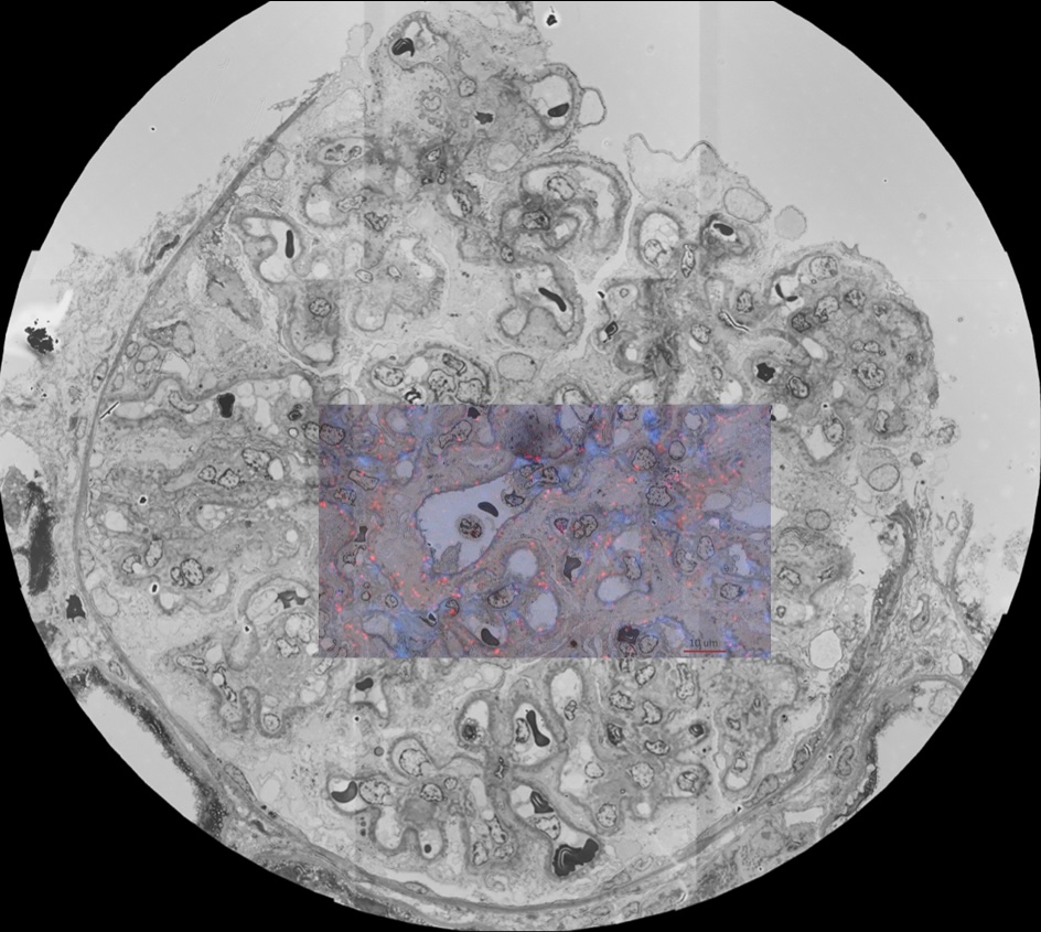
The team then re-imagined the Electron microscope. They redesigned one previously used for engineering and geology for application in pathology. A collaboration with ATA Scientific and Thermo Fisher Scientific International, it is a new class of EM capable of resolving single proteins, viruses and key cellular changes in renal disease, cancer and rare diseases, promising wide-ranging health and economic benefits for patients and the health system.
In a world-first trial, a prototype instrument was produced and assessed in NSWHP Liverpool, with results presented at the 20th International Microscopy Congress (IMC-20) in Korea in 2023. Based on these results, a second prototype is now being developed by Thermo Fisher in the UK to provide the enhancements essential to replace existing EM technology currently in use.
What is the broader implication of the research?
The project creates the possibility of a new diagnostic and research laboratory model that leverages revolutionary digital imaging technology and nanotechnology. It builds relationships with key industrial stakeholders with the technical and engineering skills to translate pathology’s vision for the instrument and the powerful microscopic resolution required.